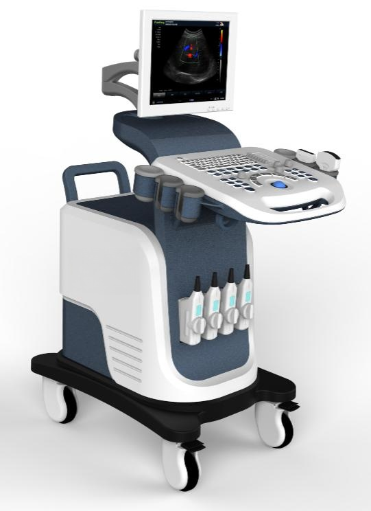
Color Doppler Ultrasonic Diagnosis System S7500

Parameters:
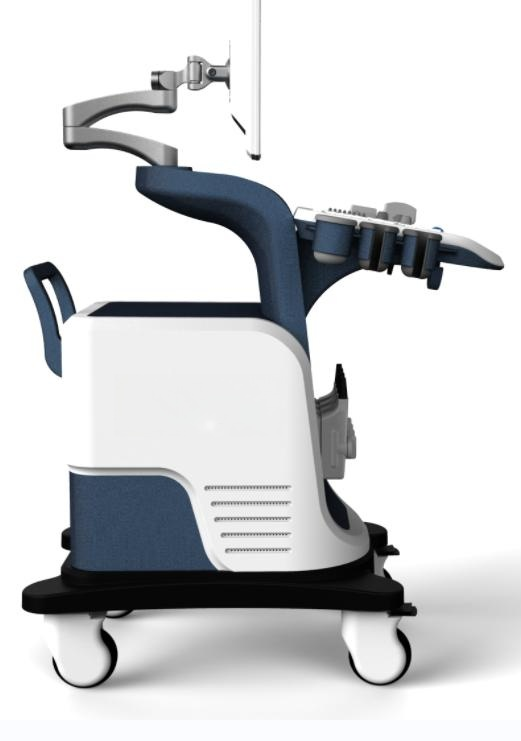
15”HD LED Displayer. Ergonomic design, All-round display angle adjustment
Ultra-wideband imaging/THI /Trapezoidal imaging/ Exact SAR adaptive speckle
noise suppression techniques.
CFM mode /PDImode / PW, Real time triplex Imaging.
6 differentlanguages System.
Rich measurement package which can satisfy clinicalneeds.
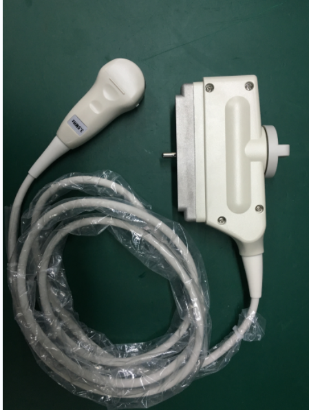
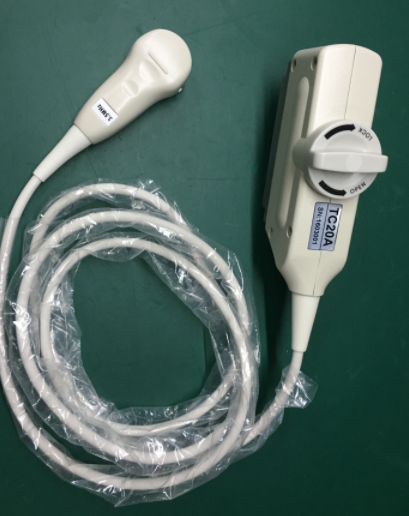
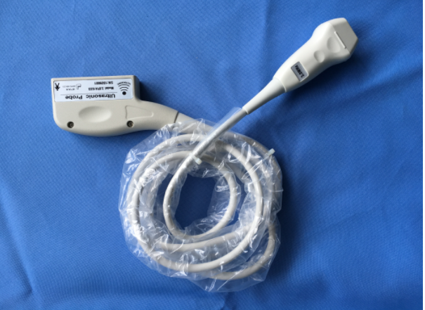
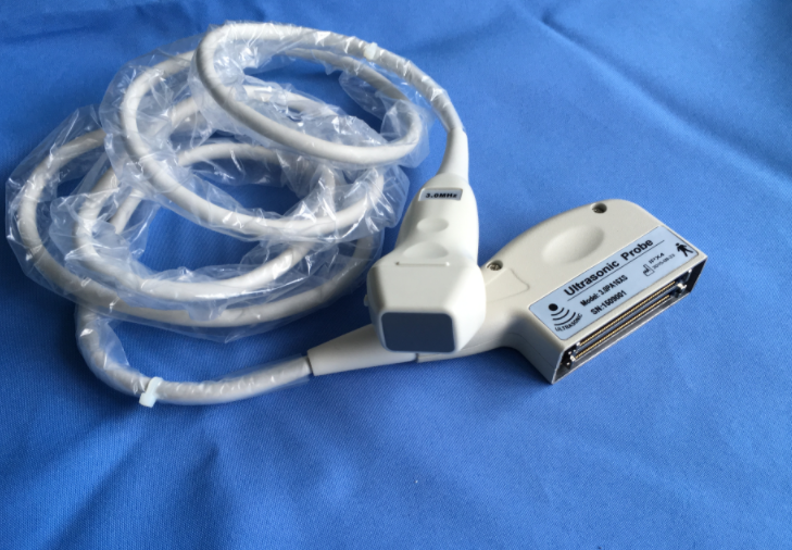
support convex, linear, trans-vaginal, micro-convex, phased array and 4D probes.
3D/4D Optinal function.
One-click optimization function.
Image and video one-click fast storage
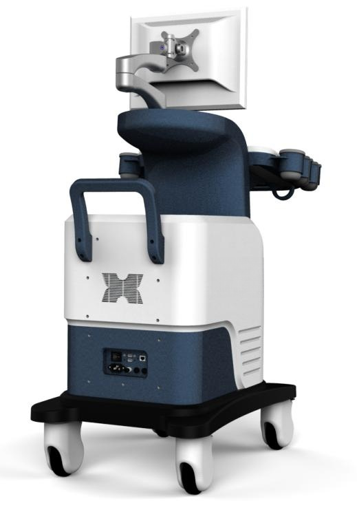
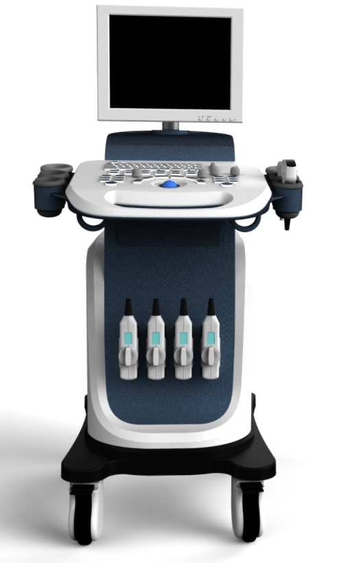
Host Parts specification table
NO | Designation | Function | Remarks |
1 | Liquid crystal display | Ultrasound images and the application of module interface display, etc |
|
2 | The probe holder | Placed temporarily does not use the probe |
|
3 | Hook drive | Probe cable hook |
|
4 | The probe socket | The probe with the host interface | Three probe socket |
5 | The monitor arm | Support the LCD display |
|
6 | Control panel | For text annotation, report writing and application of ultrasonic control operation |
|
7 | Equipment armrest | The armrest of mobile devices |
|
8 | Power switch | The device on/off power supply |
|
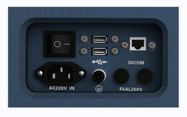
9 | The power interface board | Power supply, protective earthing, USB, DICOM interface |
|
10 | Trundle | Mobile/fixed equipment |
Main Functions | |
Scanning mode | Convex array, lumen, high-frequency linear array, phased array |
Element | 128 |
PC platform | WIN XP |
Standard probe | 2.0/2.5/3.5/4.5/5.5MHz convex |
Option probe | 2.0/3.0/3.5/4.0MHz phased array transducer 5.0/7.0/8.5/10/12MHz linear transducer 4.0/5.0/6.0/7.0/8.0MHz transvaginal transducer 4.5MHz 4D volume transducer |
Dynamic range | B、B/B、M、B/M、CFM、CMF/B、PDI、B/PW, total eight mode,( optional 4D) |
Application mode | abdomen, gynecology, obstetrics, superficial organ, urologist, heart and user defined model 1-4, total ten models |
Image mode | digital beam forming, tissue harmonic imaging |
Acoustic output | Mechanical index and thermal index real-time display |
Acoustic power | Step is adjustable, real-time display |
Gray scale | 256 scales |
Depth display | ≥250mm |
B/D dual-purpose | linear array: B/PWD; convex array: B/PWD |
Pseudo color processing | 16 kinds of pseudo color encoding can optional |
Gain adjusts | 8 segments TGC, B/M/D/C is independently adjustable; TGC curve can show and hide automatically |
Image magnification | picture in picture zoom in and zoom part function |
Image processing | Edge enhancement: Multilevel adjustable Frame average: Multilevel adjustable Line average: Multilevel adjustable Focus Optimization: Multilevel adjustable Gray Restrain: Multilevel adjustable Gamma correction: Multilevel adjustable Contrast: Adjustable Brightness: Adjustable |
Self-motion optimize function | Built-in multiple check type, according to different inspection organs, preset best image check condition, reduce the adjusting operation keys |
One-click optimization function | preset several parameters adjusting focus on a button, a key to realize image fast optimization |
Measurement and calculation | B mode routine measurement: Distance, circumference, area, volume, angle, ratio, and stenos rate. M mode routine measurement: Heart rate, time, distance, speed, ratio, etc. Gynecology measurement: Uterus, cervix, endometrial, ovary, follicular. Obstetrics measurement: EGA, ETD, fetal weight estimation, AFI index, OB report (including OB tables). Cardiology measurement: LV measurement. Urology measurement: Prostate volume, displacement volume, bladder capacity, and residual urine output. PW measurements: Time, speed, Heart Rate, RI, PI, etc. Other measurement: Slice volume measurement, hip joint angle measurement. |
Image storage | Image storage, video storage, cine loop, disk storage capacity≥128G SSD |
Patient data | Medical record management, report inquiry and printing, image video output( HDD 、USB、Optional DVD-RW)、built-in ultrasound workstation |
Reporting system | automatic report generation system, and can be full screen characters in both Chinese and English editor; |
Output interface | USB、DICOM interface |
Optional | 1.3D/4D imaging technology 2. Sound velocity imaging technology optimization 3.Color Doppler Energy(CDE) 4.Pulse Wave Doppler imaging(PW) 5.Continuous Wave Doppler Imaging(CW) 6.Color Doppler resolution enhancement 7.Panoramic imaging,space compound imaging 8.Adaptive speckle noise suppression techniques 9.Ergonomic design,All-round display angle adjustment 10.Imaging optimization technology;Compound enhance technology 11.Imaging processing technology 11.Multi-beam parallel processing technology 12.Tissue harmonic imaging 13.Four activated ports,15/17'HR medical monitor 14.One Touch Optimize under different modes |